ABOUT vMic
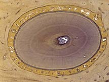
Slide acquisition was done by Dieter Glatz with a robot-microscope which scans the slide and captures a matrix of images.
For this a 40x Zeiss Apochromat with NA=0.95 has been used in order to obtain high quality images. The images were postprocessed and then served by a plain http-server. The viewer - called vMic - has been developed with Macromedia Flash.
For further information see: Human Pathology - Glatz, Glatz, Mihatsch - Volume 34, No. 10 (October 2003) - pages 968-974: Virtual Slide: high quality demand, physical limitations and affordability.
Katharina and Dieter Glatz-Krieger have developed idea and concept of the vMic.
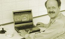
Dieter has realized the technical infrastructure and Katharina is responsible for content.
Oliver Walthard is accountable for the design of the vMic-website.
|


